Medial knee pain can emerge from multiple anatomical structures, including ligaments, menisci, bursae, and cartilage. These structures have distinct pathological mechanisms and rehabilitation demands. The variability of structures means that there are multiple potential pain generators, which require specific diagnosis. Misidentification of the underlying pathology may lead to persistent pain and disability, failed rehabilitation and unnecessary time spent away from meaningful activities.
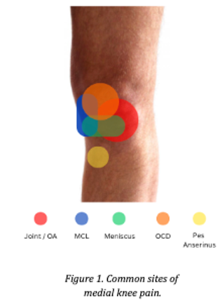
Accurate diagnosis requires a thorough understanding of one’s pain patterns, loading history, injury mechanism, and, for adolescence, growth and fuelling status. A thorough physical assessment is crucial to differentiate these structures, with additional imaging in some cases being crucial to ascertain a definitive diagnosis.
Anatomy of the Medial Knee
The medial knee comprises ligamentous restraints, meniscal structures, bursae, and cartilage surfaces, each with distinct roles in load sharing and stability.
KEY STRUCTURES
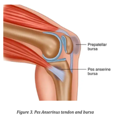
The medial collateral ligament (MCL), is the primary restraint to valgus stress. This ligament spans from the medial femoral epicondyle to the medial tibia. The medial meniscus, a C-shaped structure made of cartilage, distributes load across the medial tibiofemoral compartment and acts as a shock absorber. The pes anserinus (goose’s foot), is a conjoined tendon of the sartorius, gracilis, and semitendinosus tendons, which inserts on the anteromedial tibia, with an underlying bursa present to reduce friction between tendon and bone. The medial patellofemoral ligament (MPFL) prevents lateral patellar translation and may be disrupted during patellar dislocations. The medial tibiofemoral articulation is a common site of osteoarthritic change, a joint condition where the cartilage within the knee gradually reduces and changes shape over time.² In this area, we also have cartilage lining the ends of the femur (thigh bone), which can be susceptible to both trauma and overload, particular in adolescence.
DIFFERENTIAL DIAGNOSIS: PATHOANATOMY AND REHABILITATION PATHWAYS
MEDIAL COLLATERAL LIGAMENT INJURY
MCL injury can result from direct trauma to the lateral knee, or, valgus stress with knee flexion and external rotation. These would present with acute medial knee pain, anteromedial swelling, and positive valgus stress testing. Rehabilitation, depending on the severity of injury, may involve time away from sport, with a range of motion (ROM) hinged brace considered early on in moderate grade injuries to aid with appropriate tissue healing.1 Progressive resistive training can be performed, in spite of the knee brace. Return to sport timeframes vary depending on grade. Biology is considered, that is, allowing adequate time for the tissue to heal. However, progression through phases of rehabilitation should be criteria based, including ROM, strength, power and tolerance to both straight line and multidirectional running.

MEDIAL MENISCUS INJURY
Meniscal tears can be either degenerative (occur over time), or, occur via acute trauma. There are different types of meniscus tears, which are generally described based on their appearance and location within the meniscus. The meniscus is mostly avascular (low blood supply), with vascularation being high along the periphery, but almost absent within the central regions.2 This means that its capacity to heal is variable. Presentation may include sharp medial joint line pain, mechanical locking or catching, feelings of giving way or instability, and an effusion (swelling). Management is dependent on the type of tear, and symptoms being experienced, and can include nonoperative or operative management. Operative management is generally considered when symptoms are highly disabling, and if there is a significant lesion that affects the load bearing capacity of the tissue.
PES ANSERINUS TENDINOPATHY AND BURSITIS
The pes anserinus is a conjoined tendon made up of 3 tendons (sartorius, gracilis and semitendinosus). It assists with knee flexion, internal tibial rotation and also acts as a dynamic, secondary stabiliser for the knee joint.3 Underneath the tendon lives a small fluid sac called a bursa, which serves to provide a smooth gliding surface for the overlying tendon. With sudden spikes in activity levels, particularly hip flexion based activities, the overlying tendon may become irritated, and/or the underlying bursa may become inflamed. Pain localises to the anteromedial tibia, 5–7 cm below the joint line, and can be reproduced by resisted knee flexion and hip adduction. Some may also feel discomfort with activities like lunging and squatting. Rehabilitation generally considers managing load - that is, slowing things down and gradually building back up. Considerations can be made to strength from the hips down to the foot, as well as running mechanics.
OSTEOARTHRITIS
Osteoarthritis is a whole joint condition encompassing progressive cartilage loss and bone remodelling within the medial tibiofemoral compartment. This is an inherently normal occurrence, however, the rate at which this process occurs can be accelerated for a number of reasons. Risk factors include prior meniscal injury, ligamentous laxity, and previous surgical procedures to the knee. Its presentation is varied, however, some common symptoms include: morning stiffness (generally no longer than 30 mins), activity-related pain, stiffness after inactivity and variable swelling. Rehabilitation focuses on symptom reduction while maintaining and improving functional capacity with activity modification. Additionally, consideration to lifestyle factors is crucial. Think of the knee's capacity as a bucket. Improving strength and functional capacity may serve to increase the size of that bucket. The ‘water’ filling it up includes systemic inflammation, poor sleep, high stress, and metabolic health. If you only build strength (enlarge the bucket slightly) but neglect the elements filling the bucket, the bucket will continue to overflow, and symptoms may persist.4
OSTEOCHONDRAL DEFECT (OCD)
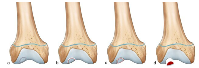
Osteochondral lesions involve damage to both the cartilage and underlying bone. Acute onset typically follows direct trauma or an impaction injury. Alternatively, these lesions can manifest over time, without being symptomatic, or, being misdiagnosed. This is particularly common in adolescence, when the cartilage is vulnerable to overload and ultimately fails due to repetitive, high stress activities. Presentation may include medial knee pain, mechanical symptoms of locking, catching or giving way and an effusion. Pain is typically worse with activity.
These lesions are generally graded by size and stability, with large, unstable lesions requiring surgical intervention.5 Stable lesions are generally managed conservatively with a period of offloading, followed by a rebuild into strength, impact and sport related activities.
Imaging is crucial to determine the status of the lesion, with MRI being the gold standard.
CLINICAL INTEGRATION
Medial knee pain can reflect multiple pathological processes concurrently. For example, MCL insufficiency secondary to meniscal tear, or referred pain from adductor pathology. Systematic evaluation enables accurate classification. Across all medial knee conditions, common rehabilitation principles emerge: local strength and proximal control may reduce medial knee stress. Lower-limb strength symmetry and mechanics are prerequisites for return to sport, and progression should be criterion-based rather than timeline-based. While specific exercise progressions differ between conditions, these underlying principles remain consistent.
CONCLUSION
Medial knee pain demands accurate diagnosis and individualised rehabilitation targeting the specific pathological process. The majority of presentations respond favourably to systematic, evidence-informed rehabilitation. When conservative management is inadequate, surgical consultation is appropriate. Athletes with medial knee pain benefit from consultation with physiotherapists to ensure accurate diagnosis and criterion-based progression toward pain-free function and sport performance.
REFERENCES
- Tandoğan NR, Kayaalp A. Surgical treatment of medial knee ligament injuries: current indications and techniques. EFORT Open Reviews. 2016;1:27 - 33. doi:10.1302/2058-5241.1.000007
- Orellana F, Grassi A, Hlushchuk R, et al. Revealing the complexity of meniscus microvasculature through 3D visualization and analysis. Scientific Reports. 2024;14. doi:10.1038/s41598-024-61497-2
- Curtis B, Huang BK, Pathria M, Resnick D, Smitaman E. Pes Anserinus: Anatomy and Pathology of Native and Harvested Tendons. AJR. American journal of roentgenology. 2019. doi:10.2214/ajr.19.21315
- Herrero-Beaumont G, Castro-Dominguez F, Migliore A, Naredo E, Largo R, Reginster JY. Systemic osteoarthritis: the difficulty of categorically naming a continuous condition. Aging Clin Exp Res. 2024;36(1):45. Published 2024 Feb 20. doi:10.1007/s40520-024-02714-w
- Salzmann G, Niemeyer P, Hochrein A, Stoddart M, Angele P. Articular Cartilage Repair of the Knee in Children and Adolescents. Orthopaedic Journal of Sports Medicine. 2018;6. doi:10.1177/2325967118760190